3D Cone-Beam Computed Tomography (CBCT) Imaging
In order to visualize and measure the upper neck alignment in a way that allows us to have pinpoint accuracy, we utilize the most state-of the-art 3D imaging for our patients. We are the the only chiropractic office in San Ramon to utilize this technology in patient care.
Unlike traditional X-RAYs that take multiple 2-D images to attempt to view the 3-D human structure, the Cone Beam computed tomography (CBCT) is a specialized type of X-RAY that can view the Cranio-Cervical Junction (CCJ) in 3 Dimensions (3-D). The imaging beam is a ‘cone-like’ formation that is moved around the patient to produce the highest quality images of bone. It is able to capture a wide variety of angles in a single scan.
Here are some answers to common questions that patients have:
What are the benefits of 3-D imaging?
- More accurate diagnosis
- Less Radiation
- Better image quality
- Wider variety of viewing angles
- Easier patient experience
- More precise treatment planning
Why is viewing the upper neck in 3-D so important?
Since the human structure is 3 dimensional, it is vital to view it with 3-D imaging in order to accurately evaluate the alignment. Traditional X-ray is 2-Dimensional, so certain factors like anatomical variations, positioning errors, and asymmetry of structures can result in misdiagnosis some of the time. For a long time, traditional X-ray was simply the best form of imaging available for the spine; that is, until we began Cone Beam Imaging.
How does the radiation exposure compare to that of typical X-ray?
The radiation exposure is much less than standard XRAY. Standard scan is 237 microsieverts, equivalent to about 1/10 the radiation exposure of a standard 4-view Cervical spine X-ray series. This is the equivalent to the exposure of a 5 hour airplane flight. We feel strongly that the small additional risk from radiation exposure is much less than the risk of inaccurate diagnosis or treatment from proceeding with the knowledge provided by a CBCT scan.
How does the 3-D imaging change expected clinical results?
Because of the increased information provided by the 3-D imaging, treatment planning is expected to be more precise and results are expected to be even greater than with traditional imaging . With the use of software, Dr. Wang can manipulate and measure the images in order to view the upper neck anatomy and surrounding structures with incredible clarity.
How long does the imaging take?
20 seconds
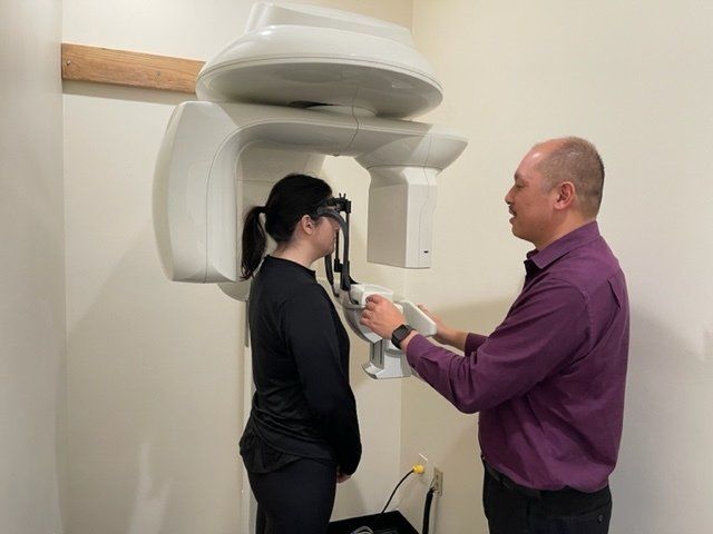
How does the procedure work?
The patient is standing with the head in a supported position. You will be asked to remain very still while the arm of the unit rotates around your head by 360 degrees, capturing multiple images from every angle. This experience is very similar to a panoramic dental X-ray.New Paragraph